In this post, we list imaging tests that assist OBS/GYNs, physicians and other doctors who offer gynecology services in assessing, diagnosing and treating patients.
What are gynecological imaging tests?
At Premier Imaging, we offer several gynecology tests and procedures for women with various conditions and health requirements. These important medical screenings can help your doctor assess your health and may help to determine whether you require gynecologic surgery or other forms of treatment.
These tests and exams include:
Pelvic Ultrasound
A gynecological pelvic ultrasound consists of both a transabdominal (on the abdomen) and a transvaginal exam, where the transducer is inserted into the vagina to obtain higher resolution images of the uterus and ovaries. The intracavitary ultrasound transducer is covered in a non-latex probe cover and coated with a sterile, water-soluble lubricant.
The technologist will image the uterus, ovaries and surrounding areas to assess for any pathology such as polyps, cysts, fibroids, etc.
A pelvic ultrasound generally takes under 30 minutes to complete.
eFTS Testing
Enhanced first-trimester screening can help us detect whether your baby may potentially be at risk for specific chromosomal conditions such as Edwards Syndrome (trisomy 18) or Down Syndrome. Also known as a first-trimester combined test, the assessment involves taking an ultrasound exam and a blood test.
The test can be done earlier than most other prenatal screening tests and offers a valuable assessment of the viability, anatomy and growth of a fetus. It allows you and your healthcare team to have more time to make informed, proactive decisions about the course of your pregnancy.
This is just a screening tool, so any abnormal results will be followed up with more diagnostic tests that will enable you and your team to create a treatment and management plan for any conditions before, during and after delivery.
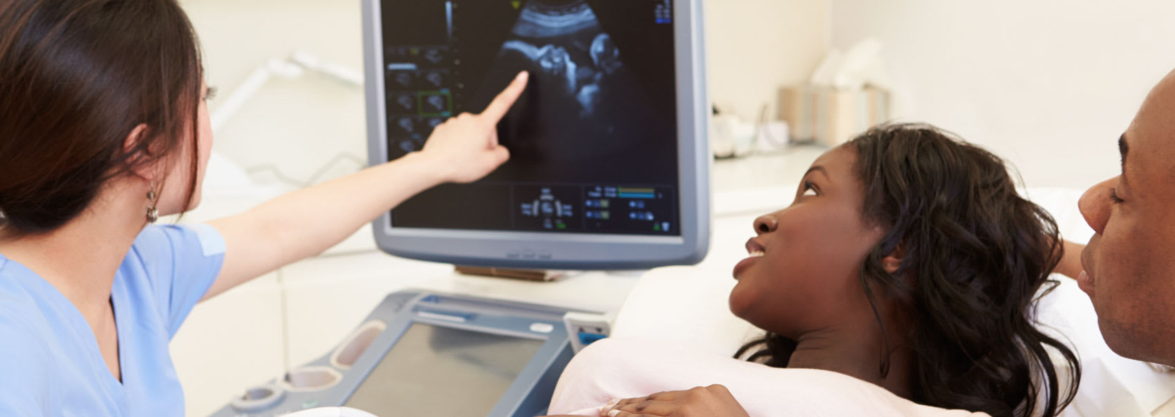
Obstetrical Ultrasound
An obstetrical ultrasound allows us to confirm pregnancy and examine the embryo or fetus in the womb of a pregnant woman. In addition, we will be able to see the ovaries and uterus.
With this test, we can establish that a living embryo or fetus is growing, estimate the length of pregnancy so far and learn other valuable information about the pregnancy and developing fetus, such as whether the fetus has congenital abnormalities.
During the test, sound waves are transmitted through the skin and an image is produced from the echoes received. The exam takes approximately half an hour but can take longer depending on the ease of obtaining all necessary images.
Saline Infusion & Tubal Patency
A saline infusion and tubal patency test can often bring women who are experiencing infertility issues accurate, clear answers. Saline infusion sonography (SIS) allows us to look into fertility problems, analyze tubal patency in infertile patients, and assess the uterine cavity.
A tubal patency study can help us answer questions around why a woman may be having challenges with conception. Saline solution is passed through the fallopian tubes so we can use ultrasound to see whether the tubes are “open” or patent.
Your doctor may also recommend SIS if you suffer from heavy, irregular or post-menopausal bleeding, or have had abnormal ultrasound results. If any further diagnostic testing is needed after this appointment, your doctor will follow up.