Pregnancy does not always feel completely real until you see your baby for the first time during your first prenatal ultrasound. These ultrasounds are a very standard — and very welcome — part of prenatal care. Today, our Ottawa radiologists describe the ultrasound process, how many you will have throughout your pregnancy, and what they can detect.
What is an ultrasound?
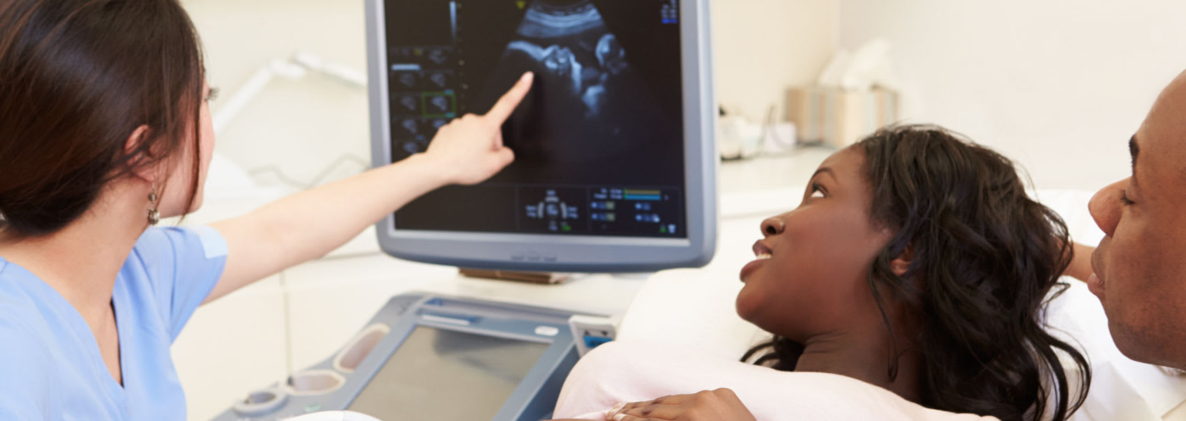
An ultrasound is a technology that creates images using sound waves. A transducer is inserted into the vagina or placed on top of the belly and emits sound waves that bounce off the baby's tissues, fluids, and bones. The transducer then detects these echoes and converts them into an image displayed on a screen.
During early pregnancy, ultrasounds confirm the fetal heartbeat and the position of the baby in the uterus. Later in pregnancy, ultrasounds examine the baby's general health and anatomy, as well as fetal growth and placenta location. At the later stages of pregnancy, ultrasounds can ensure the baby is in a head-down position before labour.
What happens at the first-trimester ultrasound?
The first ultrasound is referred to as a "dating" or "viability" ultrasound. It's usually done between 7 and 9 weeks to confirm your due date, look for a fetal heartbeat, and measure the baby's length from "crown to rump."
If your periods are irregular or you haven't had a period since stopping birth control, this ultrasound will be especially useful in determining a more accurate due date. Your due date is significant because it informs your doctor about your baby's development each month.
This first ultrasound is normally performed transabdominally or transvaginally. People may be asked to arrive with a full bladder to help position their uterus better for the ultrasound. A transvaginal exam is sometimes required to obtain a clearer view in order to date the pregnancy.
During a transvaginal ultrasound, a diagnostic medical sonographer will insert a narrow ultrasound wand just inside the vagina. The wand is covered in a non-latex probe sheath and covered with sterile gel. The wand will not enter the cervix and is therefore safe for the baby.
What happens at the second-trimester ultrasound?
The second-trimester ultrasound is also called the morphology ultrasound and is typically the one people look forward to the most. It is typically performed around the 5-month mark.
The full-anatomy scan is typically a transabdominal ultrasound. The sonographer will apply ultrasound gel to the abdomen and use the transducer to assess and capture images of the fetus
The sonographer will use tricks like asking you to lie on one side and then the other, emptying or filling the bladder, or even moving around to encourage the baby to change positions. They will do what is necessary to get the images required to track the baby's growth and development.
Your sonographer will capture the following images and measurements:
- limbs: arms, legs, feet, hands
- torso: chest, heart, kidneys, stomach, bladder, diaphragm
- head, brain structures, and face
- spine
- umbilical cord
- amount of amniotic fluid
- location, size, and shape of your placenta
- length of your cervix
The sonographer and the radiologist examine the images and measurements for anomalies like congenital heart defects or cleft lip/palate. They will send their findings to your referring physician who will go over the results with you.
This is also the time when you can find out if your baby will be a boy or a girl. If you would like to know you can inform your sonographer at the time of your appointment.
Are there additional ultrasounds during pregnancy?
Sometimes, an additional ultrasound or two might be required. For example, if your cervix is shorter than expected, you may need an additional ultrasound to check cervical length. You may need to have it checked on a regular basis to ensure that it remains closed throughout your pregnancy.
Additionally, regular ultrasounds may be performed if there are any placental abnormalities. Because the placenta is in charge of delivering blood and nutrients to the baby, it is critical to monitor its development.
Finally, if the uterus has not grown correctly in the previous months, your doctor may recommend regular growth ultrasounds. Growth ultrasounds are used to monitor a baby's size and development. They are usually completed in the late second trimester or within the third trimester of pregnancy
FAQs Regarding Ultrasounds
-
When is the first ultrasound done during pregnancy?
The first ultrasound is typically performed around 7-9 weeks of pregnancy. However, the first ultrasound can usually take place anytime during the first trimester, which normally translates to the first 13 weeks of pregnancy.
If your pregnancy is considered high-risk for any reason, it's best to have your first ultrasound sooner rather than later.
-
How soon can pregnancy be detected by ultrasound?
An ultrasound scan can detect a healthy pregnancy within the uterine cavity as early as 17 days after the egg was released from the ovary (ovulation). This occurs three days after a missed period.
However, knowing, or even suspecting, you're pregnant at this stage is extremely rare. Typically, your first ultrasound will occur at the 7-8 week mark.
-
How can I read pregnancy ultrasound abbreviations?
The following is a list of the most common abbreviations used on an ultrasound report, and their meanings:
- Biparietal diameter (BPD) - measures across the head
- Head Circumference (HC) - measures around the head
- Abdominal Circumference (AC) - measures around the abdomen
- Femur Length (FL) - measures the length of the thigh bone
Additionally, you may see SGA (small for gestational age) and LGA (large for gestational age).
Most fetuses that are small for gestational age are healthy, but some may require additional ultrasound evaluation to ensure that they are growing properly. Other ultrasound tests, such as checking the amount of amniotic fluid around the fetus and measuring blood flow in the umbilical cord (umbilical artery Doppler), can also be performed.
The majority of fetuses that are large for gestational age on ultrasounds are well nourished and healthy at birth. In some cases, the fetus's size is due to an underlying cause, such as diabetes or a genetic syndrome.
-
How many ultrasounds do you need during pregnancy?
Most women receive three ultrasound scans during pregnancy. The first is, ideally, in the first trimester to confirm the due date. The second ultrasound is around 12 weeks to measure the nuchal translucency for genetic screening purposes. This test is usually accompanied by blood work. Finally, the third ultrasound is around the 5-month mark for the assessment of the fetal anatomy.
However, there may be some situations that call for more ultrasounds. such as ones for monitoring baby’s growth.
Generally, you might need extra ultrasounds if you have:
- hypertension
- diabetes
- high BMI (body mass index) going into pregnancy
- preeclampsia
- indicators that your placenta or uterus is not growing appropriately
- advanced maternal age
-
Are ultrasounds safe during pregnancy?
Ultrasounds have been used by providers for more than 30 years, and no dangerous risks have been discovered.
When performed by your health care provider, ultrasounds are safe for both you and your baby. Ultrasounds are safer than X-rays because it uses sound waves rather than radiation.