
Our expert radiology team in Ottawa uses pelvic ultrasounds to provide a closer look at organs such as the bladder, ovaries, prostate, and uterus.

A pelvic ultrasound is a non-invasive imaging test that provides a detailed view of the organs in your pelvic area—the region between your stomach and legs. These organs include the bladder, uterus, ovaries, fallopian tubes, vagina, prostate, and rectum.
Pelvic ultrasounds are often recommended if someone is experiencing difficulty conceiving, pain during sex or urination, abdominal swelling, unusual menstrual cycles, or urinary incontinence.
They can help detect a variety of conditions, including bladder stones, hernias, and bladder cancer. Pelvic ultrasounds may also be used during certain procedures, such as biopsies, or to confirm the placement of an IUD.
The process usually starts with a short preparation, which may include drinking water to ensure your bladder is full, as this helps improve the images.
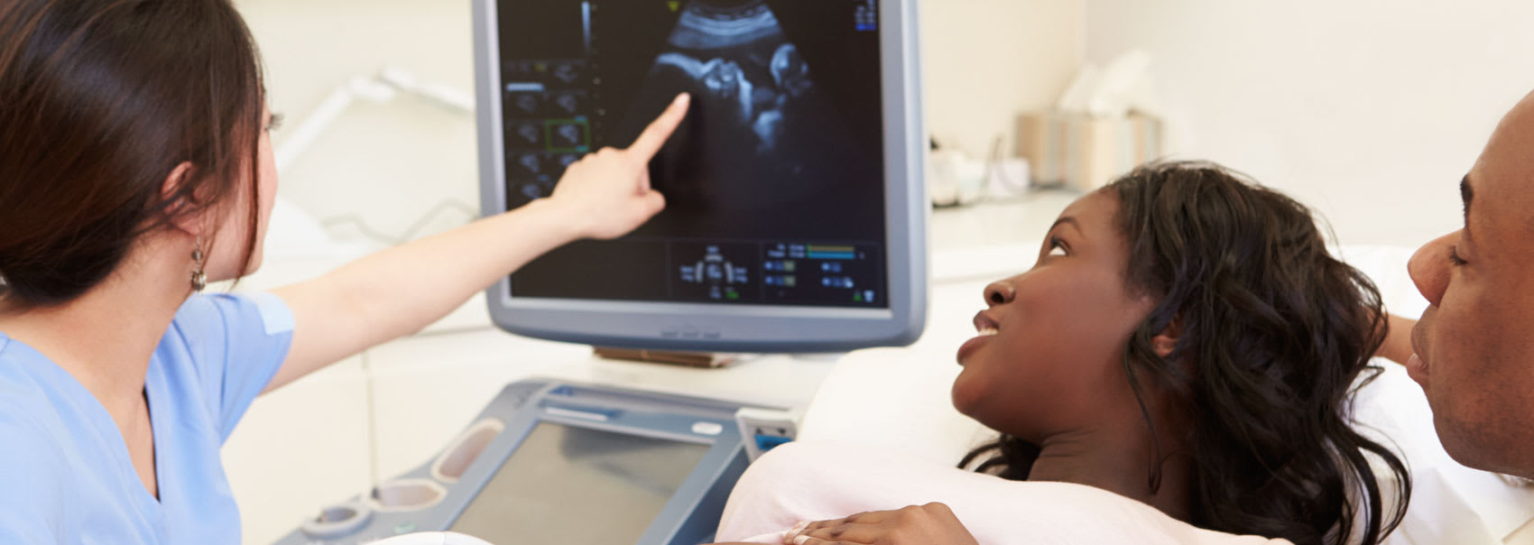
During the scan, a sonographer applies a small amount of gel to your lower abdomen and moves a handheld device called a transducer over the area to capture images. In some cases, a transvaginal or transrectal approach may be used for a closer view of certain structures.
The procedure is typically painless, takes about 15–30 minutes, and the results are usually reviewed by a radiologist who provides a report to your healthcare provider.
Here is what you can expect during each phase of the abdominal ultrasound process, along with details on what to bring with you to your appointment at our clinic in Orléans, Ottawa.
Before your pelvic ultrasound, it’s important to follow any instructions provided by your healthcare team. For some types of scans, such as a pelvic ultrasound (transabdominal), you may be asked to drink water beforehand to ensure your bladder is full, which helps produce clearer images.
Be sure to share your medical history, including any medications, surgeries, or conditions that could affect the scan.
Wearing comfortable, loose-fitting clothing is recommended, and you may be asked to change into a gown for certain types of ultrasounds. You will be given specific information on how to prepare when the exam is scheduled.
During the test, a small amount of gel is applied to your lower abdomen, and a sonographer uses a handheld device called a transducer to capture images.
For transvaginal ultrasounds, a specialized probe is gently inserted into the vagina or rectum to provide a closer view of the pelvic organs.
While you may feel slight pressure, the procedure is generally painless. Most pelvic ultrasounds take about 15 to 30 minutes to complete.
The gel will be removed from your skin once the exam is complete, but any gel that is left over will quickly dry and usually doesn't discolour or stain clothing. You should be able to return to regular activities after an pelvic ultrasound.
The images will be analyzed and interpreted by a radiologist. The radiologist will provide the doctor who requested the exam with a signed report. Your doctor will then discuss the findings with you.
A follow-up exam may be necessary to further evaluate a potential problem with additional views or a special imaging technique. It may also check to see if an issue has changed over time. Follow-up exams are frequently the best way to determine whether treatment is effective or whether a problem requires attention.
Read the answers to our most frequently asked questions about pelvic ultrasounds at Premier Imaging.
Pelvic ultrasounds can identify abnormalities in organs, such as masses or tumours, but further testing may be needed to confirm a diagnosis.
Pelvic ultrasounds are generally painless. You may feel slight pressure when the transducer is pressed against your abdomen or inserted vaginally/rectally, but it should not cause significant discomfort.
Most pelvic ultrasounds take between 15 and 30 minutes.
Preparation depends on the type of ultrasound. For a transabdominal pelvic ultrasound, a full bladder is required (drink 1–2 bottles of 500 mL of water one hour prior to your appointment). For a transvaginal ultrasound, an empty bladder is required. Your healthcare provider will give specific instructions.
Yes. Pelvic ultrasounds are non-invasive and use sound waves rather than radiation, making them safe for most patients, including during pregnancy.
A pelvic ultrasound helps visualize organs in the pelvic area, such as the bladder, uterus, ovaries, and prostate. It can be used to investigate pain, swelling, abnormal bleeding, fertility concerns, urinary problems, or to guide certain procedures like biopsies or IUD placement.
A radiologist reviews the images and prepares a report for your healthcare provider. Your provider will discuss the results with you, often at your next appointment.
We'll collaborate with your healthcare team to create a streamlined imaging and diagnostics process. Find out how we can help.