
Our expert radiology team in Ottawa uses breast ultrasounds to help evaluate the cause of breast pain, lumps, or changes seen on a mammogram.

A breast ultrasound is a non-invasive imaging exam that uses high-frequency sound waves to create real-time pictures of the tissue inside your breast. A handheld device called a transducer sends sound waves into the breast, and the returning echoes are converted into detailed images on a screen.
Breast ultrasound is especially good at showing whether an area of concern is solid or filled with fluid, such as a simple cyst. It does not use radiation, which makes it a safe option for people who are pregnant or breastfeeding.
Breast ultrasounds are not a replacement for mammograms. Instead, the two exams work together: a mammogram is used for routine screening, while the breast ultrasound is often used to take a closer look at a specific area.
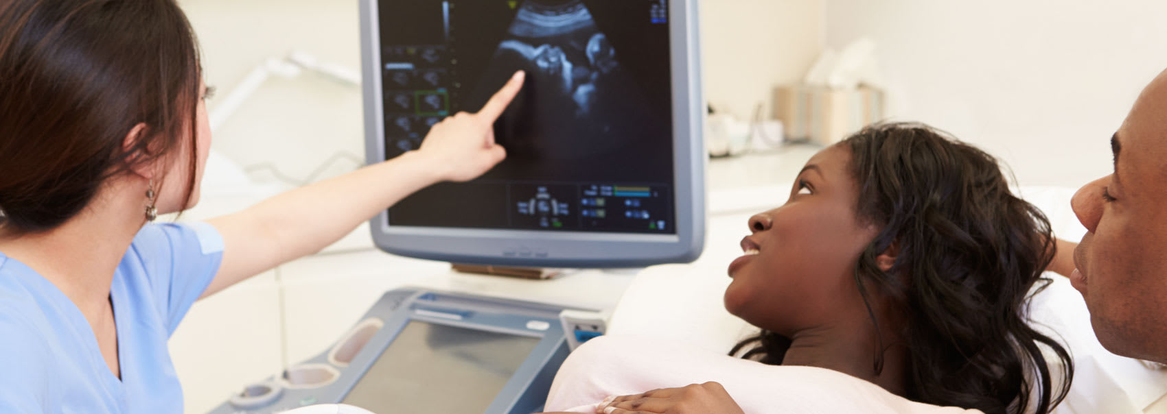
You'll lie on your back on an exam table, usually with your arm raised above your head. A clear gel is applied to the skin to help the transducer make good contact. Your sonographer then gently moves the transducer over the breast, and sometimes the underarm area, to capture images.
No special aftercare is needed, and you can return to your normal routine right away.
Your doctor may refer you for a breast ultrasound to:
Here is what you can expect during each phase of the breast ultrasound process, along with details on what to bring with you to your appointment at our clinic in Orléans, Ottawa.
No special preparation is needed. On the day of your exam:
After changing into a gown, you'll lie on the exam table, and your sonographer will apply gel to your breast. They'll move the transducer over the area being examined and may ask you to change position slightly to get the clearest images.
You may feel a light pressure, but the exam is generally painless. Most breast ultrasounds take about 15 to 30 minutes.
Any remaining gel wipes off easily and won't stain your clothes. There is no recovery time, so you can get dressed and carry on with your day.
A radiologist will review your images and send a signed report to your referring doctor, who will go over the results with you.
In some cases, and depending on your results, your doctor may recommend follow-up imaging, a short-term repeat ultrasound, or a biopsy.
Read the answers to our most frequently asked questions about breast ultrasounds at Premier Imaging.
No. Most people find it comfortable. You may feel light pressure from the transducer, especially over a tender area, but there is no compression like a mammogram.
Yes. Ultrasound uses sound waves, not radiation, so it is safe for people who are pregnant or breastfeeding.
Most breast ultrasounds take about 15 to 30 minutes, depending on how many areas need to be imaged.
An ultrasound can show changes in the breast that may need further testing, and it is very useful for telling cysts apart from solid masses. It can't confirm cancer on its own.
If something needs a closer look, your doctor may recommend a biopsy or further testing.
Yes. A breast ultrasound does not replace routine screening mammograms. Some changes, such as tiny calcium deposits, show up better in a mammogram. The two exams are often used together.
Your doctor may order one to check a lump you can feel, or because dense breast tissue can make a mammogram harder to read. An ultrasound offers another view of the same area.
Your breast ultrasound report is sent to your referring doctor once a radiologist has reviewed your images.
Your doctor will reach out to discuss the results.
We're proud to be accredited by the following organizations:


We'll collaborate with your healthcare team to create a streamlined imaging and diagnostics process. Find out how we can help.